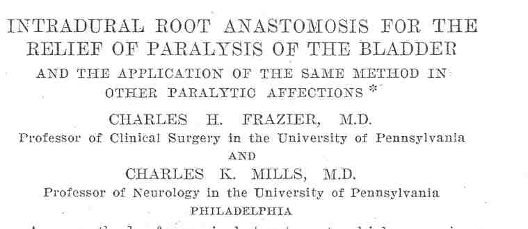
As reported in the prestigious Journal of the
American Medical Association (JAMA), U.S. scientists have for the
first time rerouted nerves from above the spinal cord injury (SCI) site to
restore some function in paralyzed areas. Specifically, Drs. Charles
Frazier and Charles Mills, University of Pennsylvania, successfully
rerouted nerves to restore bladder function in a 27-year-old man who had
sustained a lumbar-level (L2) injury after a gas tank exploded near him.
Although the patient had regained some function since
injury, his bladder remained paralyzed, and he had “absolute
incontinence.” Eight months after injury, he underwent a surgery in which
a functional L1 nerve above the injury site “was divided extradurally at
its exit from the spinal canal and brought within the dural sac” and then
sutured end-to-end to sacral-level S3 and S4-nerve roots. Eight months
later, the patient had regained some bladder control.
In previous PN articles, I discussed the
creation of function-restoring neuronal connections such as the
aforementioned. I just assumed such procedures were at the cutting-edge of
twenty-first century science. Amazingly, the surgery described
 above
was carried out in 1912!
above
was carried out in 1912!
When I came across the article (JAMA 59,
1912), I realized with dismay that function-restoring surgeries in some
form have been available for nearly a century - but relegated to the
therapeutic dustbin until recently. Given such glacial progress, it is
understandable why many people with SCI are frustrated with science’s slow
pace in producing real-world therapies.
This article will briefly review the slow emergence
of these procedures since this 1912 publication.
Another rerouting surgery was carried out in 1951 by
Dr. L. W. Freeman, Indiana University (J Neurosurg 18, 1961). In
this case, Freeman connected intercostal nerves (those leading from the
spinal cord around each rib to the sternum) to sacral nerve roots below
the injury site in a 33-year-old prisoner who sustained a thoracic (T8-9)
injury from police gunshot five months earlier. Retaining their central
spinal-cord connections, intercostal nerves were freed, routed through the
spinal canal, and connected to sacral nerve roots or implanted into the
conus medullaris (i.e., the spinal cord’s conical tip). Although the
patient believed that new leg and bladder phenomena ware attributed to the
surgery, he died four months later. Post-mortem analyses indicated the
continuity of intercostal nerve axons into both the sacral roots and
spinal cord.
Based on Freeman’s work, Dr. Hiroyasu Makino et al
(Japan) also routed intercostal nerves to paralyzed areas in eight
patients with paraplegia sustained at least a year before surgery (Neurol
Mediochir (Tokyo) 6, 1964). In four, one pair of intercostal nerves
was inserted in the conus medullaris and another pair connected to L4
nerve roots. In the other four, two pairs of intercostal nerves were
connected to L3 and L4-nerve roots. Because results were reported
relatively soon after the surgeries, only one patient at that early stage
had demonstrated significant improvement, including some ambulatory
ability.
Reported in 1980, Drs. Carl-Axel Carlsson and Torsten
Sundin (Sweden) connected thoracic T12-nerve roots to the S2 and S3-nerve
roots in two men, age 23 and 43, with L1-injuries with injuries from
accidents 10 and 14 days earlier (Spine 5(1), 1980). About a year
later, both had regained some bladder function, and one regained erectile
ability.
More recently, Dr. Shaocheng Zhang (China) has
rerouted peripheral nerves to restore function in hundreds of patients (PN,
April 2002). Restored function depended upon the specific areas that the
target nerves serve (e.g., leg muscles, bladder or bowel, etc). Zhang
often rerouted vascularized intercostal nerves, and if not long enough to
reach the target site, intervening sural-nerve segments (from the calf)
were attached. In one of Zhang’s studies using this procedure, 18 of 23
subjects regained some ambulatory function and were able to walk with
crutches or other assistive technology (Surgical Technology
International XI, 2003). Another study demonstrated that an
intercostal-sural nerve bridge restored some bladder-and-bowel function in
the majority of 30 subjects.
Dr. Giorgio Brunelli (Italy) has restored function by
redirecting the wrist’s ulnar nerve and connecting it to nerves that
control leg function. After this procedure, a patient with a complete
spinal-cord transection could stand and walk short distances. In another
procedure carried out in a woman with a complete thoracic transection, the
peroneal nerve (to the leg) was used as a bridge directly from the spinal
cord above the injury site to the nerves of the gluteus and quadriceps
muscles. After two years, she was able to walk 30-40 meters with a walker
(PN, August 2004).
Dr. Marc Tadie and colleagues (France) have rerouted
lumbar nerve roots from below the injury site to the spinal cord above the
injury site, creating a functional neuronal pathway from brain to
paralysis-affected leg muscles. This rerouting was done in a man who
sustained a complete T9-injury at age 52 three years earlier in an auto
accident (J Neurotrauma 19(2), 2002). Specifically, three
6-cm-long nerve segments from the patient were implanted on each side of
the cord at the T7-8 level immediately above the injury site. The opposite
ends were sutured to L2-4 nerve roots, which had been detached from the
point where they exit the cord. Eight months after surgery, the patient
was able to initiate some contraction of adductor and quadriceps leg
muscles, activity which was electrophysiologically confirmed.
Finally, Dr. A Livshits et al (Russia and Israel)
connected intercostal nerves above the injury site to nerve roots below
the injury site in 11 patients with complete L1-injuries (Spinal Cord
42(4), 2004). Specifically, intercostal nerves were transferred through a
vertebral canal created under deep spinal muscles and then connected
end-to-end to S2-3 nerve roots. Some bladder function was restored in all
patients.
Conclusion
Although I usually see much promise and potential on
the SCI horizon, I was dismayed to learn that function-restoring,
nerve-rerouting surgeries have existed in some form for nearly a century.
I have personally observed such surgeries and the dramatic improvements
accruing from them. Overall, I strongly believe that such procedures can
restore significant life-enhancing function in many with SCI.
Given its key role in developing new therapies, I’m
troubled that the National Institutes of Health (NIH) with its nearly
$30-billion research budget seemingly can not duplicate century-old
results while a growing number of foreign scientists are able to do so. As
a former senior NIH official, I believe this is partially the result of
NIH’s approach to funding the most scientifically meritorious of
laissez-faire submitted grant applications. Although the process
sounds good, it is based on a questionable assumption that funding the
best science automatically translates into the greatest potential for
spinning off new therapies. In fact, the result is usually that the
development of real-world therapies becomes secondary to scientific
agendas.
Although I’m in awe watching surgeons connect nerves
that I can barely see, this nerve-rerouting approach represents
theoretically a simple concept. Perhaps, this is an example of NIH overly
emphasizing sophisticated science agendas at the expense of non-glamorous,
but much more promising, surgical approaches.
Adapted from article appearing in December 2005 Paraplegia News (For subscriptions,
call 602-224-0500) or go to www.pn-magazine.com).
TOP